AE-Adult-Echocardiography Certification Test Answers, Exam AE-Adult-Echocardiography Preparation
Wiki Article
BONUS!!! Download part of TestSimulate AE-Adult-Echocardiography dumps for free: https://drive.google.com/open?id=14yGuwTf6FCX7drBFzwKdNyYyVOE4iv1P
Before the clients buy our AE-Adult-Echocardiography guide prep they can have a free download and tryout. The client can visit the website pages of our product and understand our AE-Adult-Echocardiography study materials in detail. You can see the demo, the form of the software and part of our titles. To better understand our AE-Adult-Echocardiography Preparation questions, you can also look at the details and the guarantee. So it is convenient for you to have a good understanding of our product before you decide to buy our AE-Adult-Echocardiography training materials.
Our AE-Adult-Echocardiography study questions will update frequently to guarantee that you can get enough test banks and follow the trend in the theory and the practice. That is to say, our product boosts many advantages and to gain a better understanding of our AE Adult Echocardiography Examination guide torrent. It is very worthy for you to buy our product and please trust us. If you still can’t fully believe us, please read the introduction of the features and the functions of our product as follow.
>> AE-Adult-Echocardiography Certification Test Answers <<
Exam AE-Adult-Echocardiography Preparation & AE-Adult-Echocardiography Exam Introduction
If you want to know more about our test preparations materials, you should explore the related AE-Adult-Echocardiography exam Page. You may go over our AE-Adult-Echocardiography brain dumps product formats and choose the one that suits you best. You can also avail of the free demo so that you will have an idea how convenient and effective our AE-Adult-Echocardiography exam dumps are for AE-Adult-Echocardiography certification. With TestSimulate, you will not only get a single set of copyright for AE-Adult-Echocardiography Exams but also a simulate software for real exams. Rather we offer a wide selection of copyright for all other exams under the AE-Adult-Echocardiography certification. This ensures that you will cover more topics thus increasing your chances of success. With the multiple learning modes in AE-Adult-Echocardiography practice exam software, you will surely find your pace and find your way to success.
ARDMS AE-Adult-Echocardiography Exam copyright Topics:
| Topic | Details |
|---|---|
| Topic 1 |
|
| Topic 2 |
|
| Topic 3 |
|
| Topic 4 |
|
| Topic 5 |
|
ARDMS AE Adult Echocardiography Examination Sample Questions (Q104-Q109):
NEW QUESTION # 104
Which view best demonstrates a wall thickening abnormality of the apical lateral segment?
- A. Mid-parastemal short axis
- B. Two-chamber
- C. Parasternal long axis
- D. Four-chamber
Answer: B
Explanation:
The two-chamber apical view allows visualization of the left ventricle's anterior and inferior walls, including the apical lateral segment. It is ideal for assessing wall thickness and segmental wall motion abnormalities in this region.
The four-chamber view visualizes septal and lateral walls but does not optimally display the apical lateral segment. Parasternal long axis primarily visualizes the anterior septum and posterior wall but is limited for lateral apex. The mid-parasternal short axis focuses on mid-ventricular segments and does not visualize the apex.
This anatomical and echocardiographic detail is described in the "Textbook of Clinical Echocardiography,
6e", Chapter on Left Ventricular Segmental Analysis#20:120-125Textbook of Clinical Echocardiography#.
NEW QUESTION # 105
What does the Qp represent in an atrial septal defect shunt ratio measurement (Qp/Qs)?
- A. Left ventricular outflow tract (LVOT) time velocity integral
- B. Stroke volume of the LVOT
- C. Right ventricular outflow tract (RVOT) time velocity integral
- D. Stroke volume of the RVOT
Answer: D
Explanation:
In the calculation of the shunt ratio Qp/Qs, Qp represents pulmonary blood flow, which is calculated as the stroke volume of the right ventricular outflow tract (RVOT). Stroke volume is obtained by measuring the RVOT cross-sectional area and the RVOT time velocity integral (VTI).
Qp (pulmonary flow) divided by Qs (systemic flow) quantifies the magnitude of left-to-right shunting in atrial septal defects and other congenital heart diseases.
This method is described in the "Textbook of Clinical Echocardiography, 6e", Chapter on Shunt Quantification and Flow Calculations#20:360-365Textbook of Clinical Echocardiography#.
NEW QUESTION # 106
A patient with a ventricular septal defect, an atrial septal defect, and a cleft mitral valve is likely to have which abnormality?
- A. Marfan syndrome
- B. Shone syndrome
- C. Atrioventricular canal defect
- D. Ebstein anomaly
Answer: C
Explanation:
Comprehensive and Detailed Explanation From Exact Extract:
Atrioventricular canal defect (AV canal defect) is a congenital cardiac malformation characterized by defects in the atrial and ventricular septa, along with abnormalities of the atrioventricular valves including cleft mitral valve. These features collectively cause shunting and valve regurgitation.
Ebstein anomaly primarily involves the tricuspid valve and right atrium, Marfan syndrome is a connective tissue disorder with different manifestations, and Shone syndrome involves left-sided obstructive lesions.
This is clearly outlined in the "Textbook of Clinical Echocardiography, 6e", Chapter on Congenital Heart Defects - Atrioventricular Septal Defects#20:120-125Textbook of Clinical Echocardiography#.
NEW QUESTION # 107
Which is the most likely abnormality represented in these images from a 48-year-old man with shortness of breath?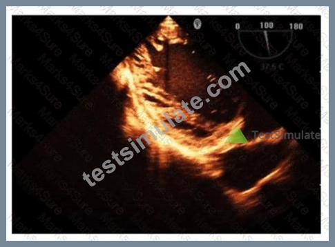
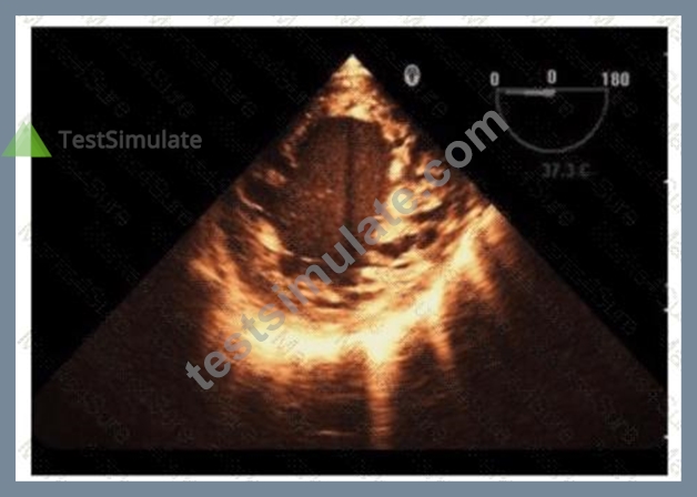
- A. Left ventricular noncompaction
- B. Loeffler syndrome
- C. Ischemic cardiomyopathy
- D. Hypertrophic cardiomyopathy
Answer: A
Explanation:
The echocardiographic images show prominent trabeculations and deep intertrabecular recesses communicating with the left ventricular cavity, best seen on contrast-enhanced images. This finding is characteristic of left ventricular noncompaction (LVNC), a cardiomyopathy resulting from arrested myocardial compaction during embryogenesis.
LVNC is diagnosed by visualizing a two-layered myocardium with a thin compacted epicardial layer and a thicker noncompacted endocardial layer with deep trabecular recesses. The use of contrast echocardiography enhances endocardial border delineation and recess visualization, increasing diagnostic accuracy.
Loeffler syndrome (hypereosinophilic cardiomyopathy) often shows endomyocardial fibrosis and restrictive physiology but not prominent trabeculations. Hypertrophic cardiomyopathy shows asymmetric septal hypertrophy without deep recesses. Ischemic cardiomyopathy shows wall motion abnormalities but not characteristic trabecular patterns.
These diagnostic criteria and imaging features are well documented in the "Textbook of Clinical Echocardiography" and ASE guidelines on cardiomyopathies and use of contrast echo#16:Textbook of Clinical Echocardiography, 6eChapter on LV Noncompaction##12:ASE Contrast Echocardiography Guidelinesp.180-190#.
NEW QUESTION # 108
Which statement is considered true regarding tricuspid annular plane systolic excursion (TAPSE)?
- A. It is an indirect measure of left ventricular systolic function.
- B. It is a measure of right ventricular diastolic function.
- C. The lower reference value is 13 mm.
- D. It is angle dependent.
Answer: C
Explanation:
TAPSE measures the longitudinal systolic excursion of the tricuspid annulus towards the apex and is a widely used echocardiographic parameter of right ventricular systolic function. It is not a measure of diastolic function nor an indirect measure of left ventricular function.
TAPSE is relatively angle independent because it is measured in M-mode from the apical four-chamber view aligned with annular motion.
The lower normal limit for TAPSE is generally accepted as 16 mm, but 13 mm is sometimes cited as a threshold below which right ventricular systolic dysfunction is suggested.
This information is presented in the "Textbook of Clinical Echocardiography, 6e", Chapter on Right Ventricular Function Assessment#20:320-325Textbook of Clinical Echocardiography
NEW QUESTION # 109
......
With the help of our AE-Adult-Echocardiography study guide, you can adjust yourself to the exam speed and stay alert according to the time-keeper that we set on our AE-Adult-Echocardiography training materials. Therefore, you can trust on our AE-Adult-Echocardiography exam materials for this effective simulation function will eventually improve your efficiency and assist you to succeed in the AE-Adult-Echocardiography Exam. And we believe you will pass the AE-Adult-Echocardiography exam just like the other people!
Exam AE-Adult-Echocardiography Preparation: https://www.testsimulate.com/AE-Adult-Echocardiography-study-materials.html
- 2026 AE-Adult-Echocardiography Certification Test Answers | Efficient ARDMS AE-Adult-Echocardiography: AE Adult Echocardiography Examination 100% Pass ???? Search for ▷ AE-Adult-Echocardiography ◁ and easily obtain a free download on ✔ www.prepawayexam.com ️✔️ ❕Reliable AE-Adult-Echocardiography Exam Materials
- Actual AE-Adult-Echocardiography Test ???? New AE-Adult-Echocardiography Exam Duration ???? Reliable AE-Adult-Echocardiography Exam Materials ???? Search for ☀ AE-Adult-Echocardiography ️☀️ and obtain a free download on ⇛ www.pdfvce.com ⇚ ????Reliable AE-Adult-Echocardiography Test Prep
- Reliable AE-Adult-Echocardiography Test Forum ???? Latest AE-Adult-Echocardiography Test Question ???? Reliable AE-Adult-Echocardiography Test Prep ⭕ Search for ( AE-Adult-Echocardiography ) and easily obtain a free download on ▷ www.examcollectionpass.com ◁ ????New AE-Adult-Echocardiography Exam Sample
- Reliable AE-Adult-Echocardiography Exam Materials ???? New AE-Adult-Echocardiography Exam Sample ♣ Top AE-Adult-Echocardiography Dumps ???? ➤ www.pdfvce.com ⮘ is best website to obtain ▷ AE-Adult-Echocardiography ◁ for free download ????Training AE-Adult-Echocardiography Material
- Pass Guaranteed Quiz 2026 AE-Adult-Echocardiography: Latest AE Adult Echocardiography Examination Certification Test Answers ???? Immediately open ▛ www.validtorrent.com ▟ and search for ➠ AE-Adult-Echocardiography ???? to obtain a free download ????AE-Adult-Echocardiography PDF Questions
- Choose The Right ARDMS AE-Adult-Echocardiography and Get Certified Today! ???? Open ➥ www.pdfvce.com ???? enter ⇛ AE-Adult-Echocardiography ⇚ and obtain a free download ????Latest AE-Adult-Echocardiography Test Question
- Pass Guaranteed Quiz 2026 AE-Adult-Echocardiography: Latest AE Adult Echocardiography Examination Certification Test Answers ???? Open ➥ www.vceengine.com ???? and search for 「 AE-Adult-Echocardiography 」 to download exam materials for free ????Reliable AE-Adult-Echocardiography Test Forum
- 2026 AE-Adult-Echocardiography Certification Test Answers | Efficient ARDMS AE-Adult-Echocardiography: AE Adult Echocardiography Examination 100% Pass ???? Download 《 AE-Adult-Echocardiography 》 for free by simply searching on ( www.pdfvce.com ) ????Test AE-Adult-Echocardiography Voucher
- Free PDF ARDMS - AE-Adult-Echocardiography - AE Adult Echocardiography Examination Unparalleled Certification Test Answers ‼ Open [ www.testkingpass.com ] and search for { AE-Adult-Echocardiography } to download exam materials for free ????Valid AE-Adult-Echocardiography Test Objectives
- Top AE-Adult-Echocardiography Dumps ???? Reliable AE-Adult-Echocardiography Exam Materials ???? Top AE-Adult-Echocardiography Dumps ???? Simply search for ▶ AE-Adult-Echocardiography ◀ for free download on ▷ www.pdfvce.com ◁ ????Test AE-Adult-Echocardiography Voucher
- ARDMS AE-Adult-Echocardiography Pre-Exam Practice Tests | www.prep4away.com ???? Open ➡ www.prep4away.com ️⬅️ enter 「 AE-Adult-Echocardiography 」 and obtain a free download ????New AE-Adult-Echocardiography Exam Sample
- keithhuyg492608.blogs100.com, www.stes.tyc.edu.tw, lexieofla941135.blgwiki.com, www.stes.tyc.edu.tw, www.stes.tyc.edu.tw, mohamadcojt640836.bloguerosa.com, nanniefzue715520.bloggosite.com, sb-bookmarking.com, roxannmbtp359096.tkzblog.com, alysharhpp191921.ssnblog.com, Disposable vapes
BTW, DOWNLOAD part of TestSimulate AE-Adult-Echocardiography dumps from Cloud Storage: https://drive.google.com/open?id=14yGuwTf6FCX7drBFzwKdNyYyVOE4iv1P
Report this wiki page